Retinal Disease Research
Find out what our researchers are doing in the area of retinal disease.

Find out what our researchers are doing in the area of retinal disease.
The Burns Lab is involved in understanding the fundamentals of retinal structure, including individual variations in normal retinal structure. We are also very interested in how retinal function and structure are influenced by disease. To do this we develop state of the art optical technology based on Adaptive Optics, and we then apply these techniques to the study of the impact of disease on retinal structure and function. 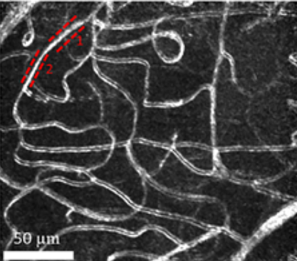
A motion contrast map of retinal capillaries (Warner 2021).
Visit the Burns Lab Website
Dr. Ann Elsner collaborates on both basic research and patient-based research to develop retinal imaging and visual function techniques to combat vision loss. Her main areas are aging, age-related macular degeneration, and diabetic retinopathy and diabetic macular edema. A main goal is to decrease the cost of eyecare by providing robust but lower cost diagnostics.
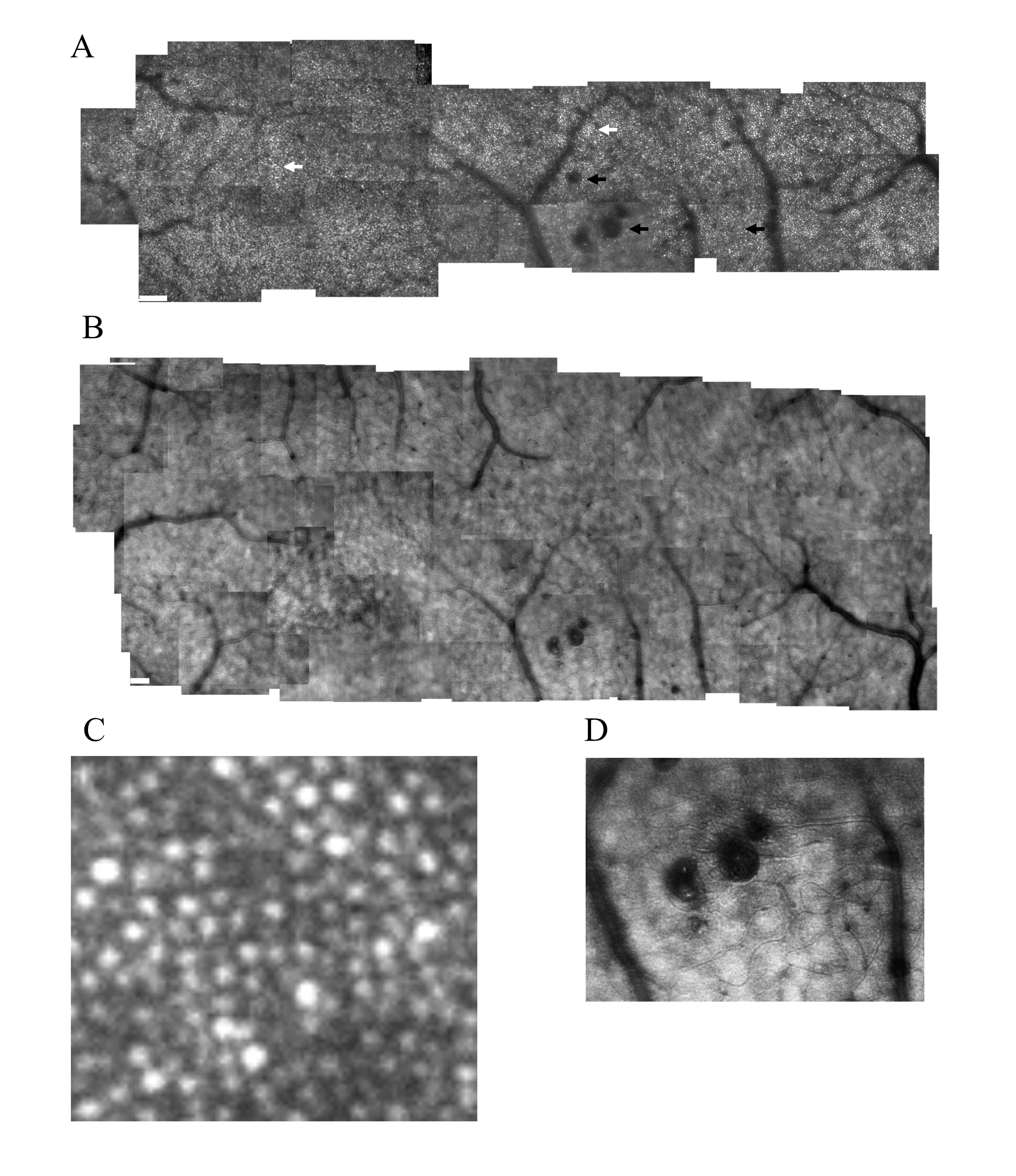
Cone distribution for a 59 yr old female diabetic subject, collected with the confocal mode of the AOSLO (A), shown in a montage of cone images with the foveal region on the left. The retinal microvasculature layer montage (B) shows a strong response to ischemia, with numerous retinal vascular changes shadowing the cone layer: multiple microaneurysms with vascular remodeling surrounding them, capillary bends or loops, and doubled or collateral vessels, particularly near microaneurysms or complex tangles. The montage of individual images shows that cone density is highest in the fovea, then decreases with increasing eccentricity, with single cones or patches of cones being unusually bright (white arrows), as well as patches of dark cones. There are regions in which the overlying retinal vessel remodeling interferes with measurements of cones, by obscuring them (top black arrow). Sometimes the contrast across an entire individual image sequence of cone images was decreased, either due to the tear film or a decreased the wavefront correction across in a large area of edema (bottom left black arrow). Elsewhere, the contrast is decreased for regions smaller than an entire image, which rules out anterior segment artifact as the sole source of image degradation (bottom right arrow). Small white scale bars at lower left = 100 microns. A magnified region of 100 x 100 microns (C) shows patches of bright cones and areas of decreased contrast. An enlargement of the retinal microvascular layer (D) shows microaneurysms and extensive vessel remodeling.
Dr. Mallika Valapala Cellular mechanisms underlying age-related macular degeneration